
Deep Learning for Pediatric Spinal Cord Injury Severity Classification
A step toward objective, imaging-based evaluation of spinal cord injury severity in children.
Summary
This study, published in the American Journal of Neuroradiology (AJNR, 2025), introduces a deep learning framework for classifying pediatric spinal cord injuries (SCI) and assessing injury severity using structural MRI measures across all vertebral levels.
By combining quantitative spinal biomarkers, cross-sectional area (CSA), anterior-posterior (AP) width, and right-left (RL) width, with a custom convolutional neural network (CNN), our model achieved:
- 96.6% accuracy distinguishing SCI from typically developing (TD) participants
- 94.9% accuracy predicting AIS (ASIA Impairment Scale) categories
This represents the first comprehensive pediatric analysis spanning C1-T11 levels, establishing MRI-derived spinal structure as a potential objective biomarker for SCI severity assessment.
Why It Matters
Assessing SCI in children is uniquely challenging. The ASIA Impairment Scale (AIS), the clinical gold standard, depends on cooperation during complex neurological testing. In young children or unresponsive patients, this can be difficult or impossible.
MRI offers an alternative: by quantifying structural changes in the spinal cord, it can objectively reflect injury severity. However, these subtle patterns are often too complex for traditional analysis, motivating our deep learning approach.
Our goal was to:
- Quantify spinal cord structural alterations in pediatric SCI versus TD controls across all vertebral levels.
- Develop a deep learning model capable of predicting AIS grades from MRI-derived structural measures.
Study Design
Participants:
61 pediatric subjects (ages 6-18), including 20 with chronic SCI and 41 typically developing controls, were scanned on a 3T MRI system.
MRI acquisition:
3D T2-weighted isotropic SPACE sequence (1×1×1 mm³ voxel size) covering C1 through T11.
Feature extraction:
Using the Spinal Cord Toolbox (SCT), we automatically segmented, labeled, and measured:
- Cross-sectional area (CSA)
- Anterior-posterior (AP) width
- Right-left (RL) width
Each metric was averaged at every vertebral level.
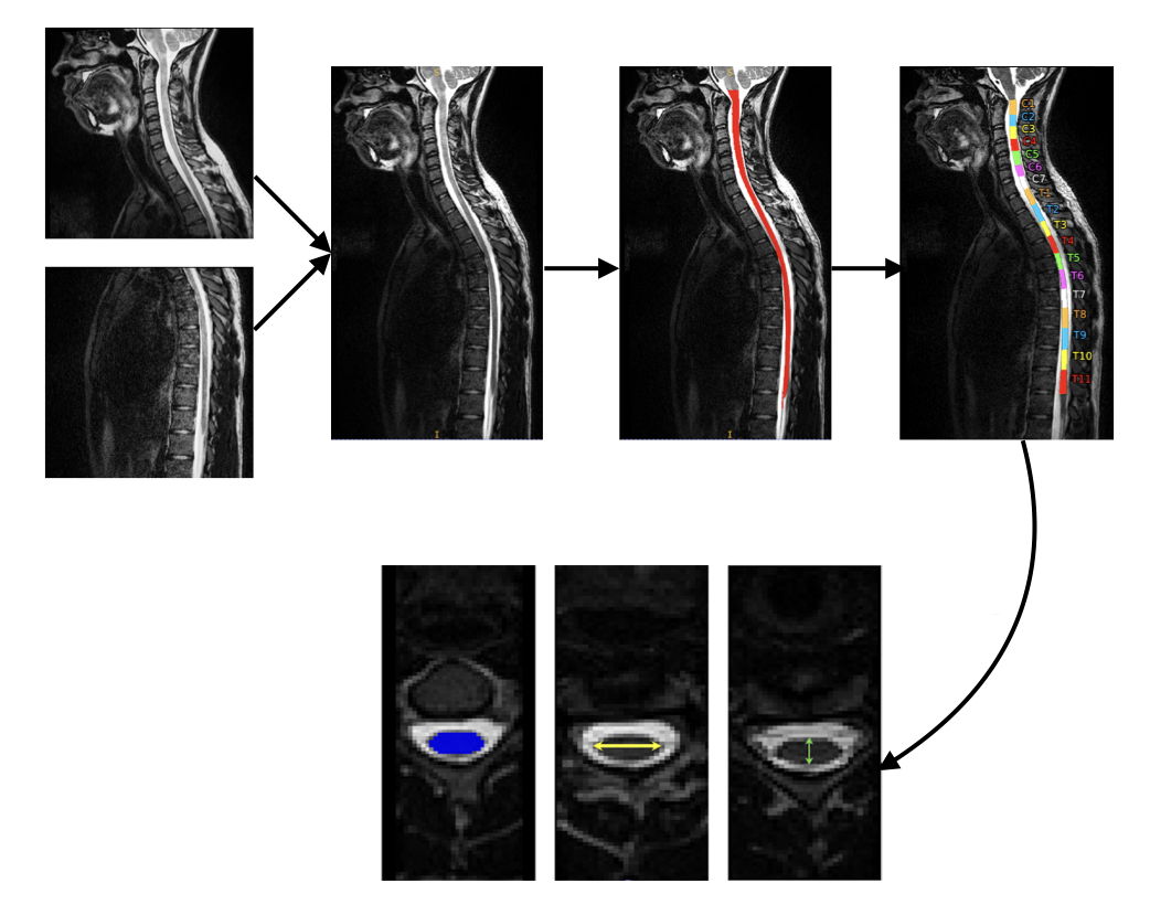
Automatic segmentation, vertebral labeling, and extraction of CSA, AP, and RL widths across all levels.
Deep Learning Model
A custom convolutional neural network (CNN) was designed to learn from structural metrics across all spinal levels.
Key design features:
- Three convolutional layers (32-64-128 filters) with ReLU activation
- Max-pooling layers and dropout (0.2) to prevent overfitting
- Integration of demographic features (age, height) in the fully connected layers
- Final softmax layer for binary classification (SCI vs TD) and multi-class AIS prediction
Training used categorical cross-entropy with the Adam optimizer (learning rate = 0.001), and early stopping to avoid overfitting. All experiments were run on an NVIDIA A100 GPU cluster.
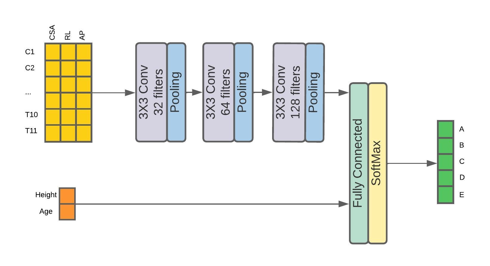
The CNN extracts spatial and structural patterns from C1-T11 measurements, integrating age and height before predicting SCI severity.
Results
Structural Analysis:
Significant differences (p < 0.05) were observed between SCI and TD participants for all measures.
- CSA: consistently smaller in SCI across all vertebral levels
- RL width: the most sensitive indicator of severity
- AP width: significantly reduced at most levels (except C2-C3, T5)
Correlation analysis showed strong relationships between MRI measures and AIS grades (up to r = 0.56 for RL width).
Model Performance:
| Model | TD/SCI Classification | AIS Classification |
|---|---|---|
| SVM | 76.4% | 68.9% |
| Random Forest | 78.4% | 74.0% |
| CNN (Proposed) | 96.6% | 94.9% |
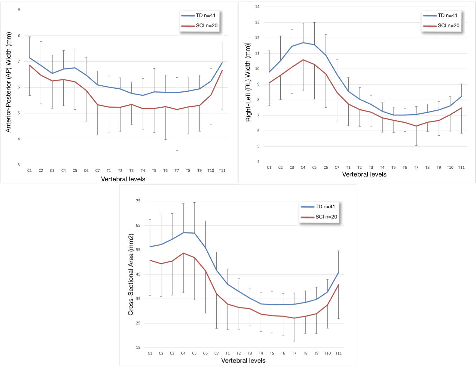
Mean CSA, RL, and AP widths across C1-T11 in TD vs SCI participants. RL width showed the strongest correlation with AIS.
Interpretation
The CNN effectively captured subtle, distributed structural changes across the spinal cord, achieving near-perfect classification, even in cases where radiologists observed no visible abnormal signal.
This demonstrates that quantitative MRI metrics can encode clinically meaningful information beyond what’s visible on standard scans, and that deep learning models can detect these patterns automatically.
Key Takeaways
- Quantitative structural MRI can objectively reflect spinal cord injury severity.
- Deep learning enables accurate, reproducible severity classification across all spinal levels.
- RL width and CSA are the strongest biomarkers linked to AIS grades.
- The framework provides a foundation for objective pediatric SCI assessment, potentially reducing reliance on subjective clinical tests.
Future Directions
While this study focused on chronic pediatric SCI, the same pipeline can be extended to:
- Acute-phase imaging to explore early predictors of recovery
- Longitudinal analysis to track structural changes over time
- Integration of diffusion MRI (DTI, NODDI) for microstructural insight
- Multi-modal models combining structural and functional MRI for comprehensive severity assessment
Citation
Sadeghi-Adl Z, Naghizadehkashani S, Middleton D, Krisa L, Alizadeh M, Flanders A E, Faro S H, Wang Z, Mohamed F B.
“Severity Classification of Pediatric Spinal Cord Injuries Using Structural MRI Measures and Deep Learning: A Comprehensive Analysis Across All Vertebral Levels.”
AJNR Am J Neuroradiol, 2025. 👉 Read the full paper