Magnetization Transfer Ratio in the Typically Developing Pediatric Spinal Cord
Establishing normative magnetization transfer ratio (MTR) values in the developing spinal cord to advance pediatric neuroimaging.
Summary
This study, published in the Journal of Neuroimaging (2025), presents the first comprehensive atlas-based evaluation of magnetization transfer ratio (MTR) values in the typically developing pediatric spinal cord (SC).
By analyzing high-resolution MRI scans from 55 children (ages 6-17), we quantified white matter (WM) and tract-specific MTR values across cervical levels (C2-C7), exploring relationships with age, sex, height, and weight.
We found that MTR increases with age, particularly at the C5 level, reflecting ongoing myelination during development. Significant sex-related differences were also observed, with females showing higher MTR in the corticospinal tracts. These normative values provide an essential baseline for evaluating pediatric spinal cord pathologies.
Background
Magnetization transfer (MT) imaging is a powerful MRI technique that measures macromolecular-bound protons, providing an indirect estimate of myelin content.
Although widely used in adults for conditions like multiple sclerosis and cervical spondylotic myelopathy, pediatric spinal cord studies are scarce.
Given that spinal cord microstructure evolves rapidly during childhood and adolescence, establishing normative MTR data is crucial to distinguish normal developmental changes from pathology.
Methods
Participants:
33 females (mean age 12.8 ± 2.8 years) and 22 males (mean age 13.9 ± 2.1 years), all neurologically healthy and screened for spinal abnormalities.
MRI Acquisition:
All scans were performed on a 3T Siemens Prisma MRI using MT-weighted fast low-angle shot sequences (FOV = 230 mm, voxel = 0.9×0.9×5 mm³, TR/TE = 35/3.1 ms).
High-resolution 3D T2-weighted images were also obtained for segmentation and registration.
Processing:
Data were analyzed using the Spinal Cord Toolbox (SCT) and the PAM50 template for tract-based alignment and quantification.
MTR values were extracted for:
- Whole white matter (WM)
- Left/right fasciculus gracilis
- Left/right fasciculus cuneatus
- Left/right lateral corticospinal tracts (LCST)
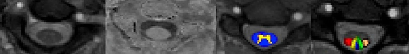
Automatic segmentation, vertebral labeling, and white/gray matter tract mapping using the PAM50 atlas.
Results
1. Normative MTR Values
Average MTR values were consistent across tracts, with low-to-moderate intersubject variability.
The LCST showed the least variability, reflecting consistent segmentation and measurement.
| Spinal Region | Mean MTR ± SD | Coefficient of Variation |
|---|---|---|
| Whole WM | 40.06 ± 2.5 | 6.2% |
| Left LCST | 40.02 ± 1.6 | 4.0% |
| Right LCST | 40.51 ± 2.5 | 6.1% |
| Left F. Gracilis | 39.79 ± 2.7 | 6.8% |
| Right F. Gracilis | 40.81 ± 2.5 | 6.1% |
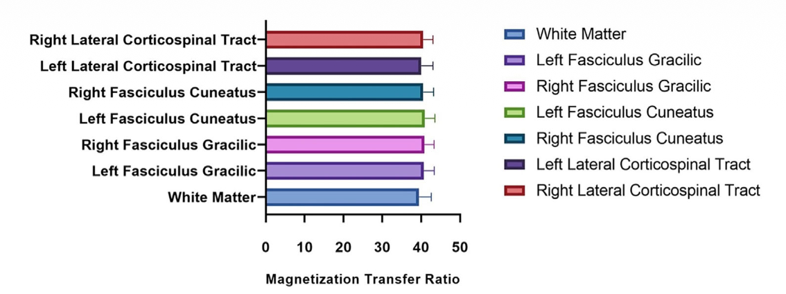
Distribution of average MTR values across cervical white matter and individual tracts (C2-C7).
2. Rostro-Caudal Differences
Analysis of variance (ANOVA) revealed significant differences (p = 0.008) in MTR across cervical levels, particularly between C2/C3 and C5, showing a decreasing trend from rostral to caudal levels.
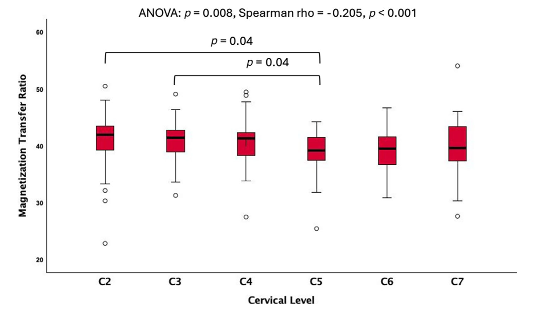
MTR values decrease gradually from upper (C2) to lower (C7) cervical levels, reflecting tract density and myelination differences.
3. Demographic Correlations
- Sex: Females showed higher MTR in LCST (+37%, p = 0.03) and total WM (+33%, p = 0.04) than males.
- Age: A significant positive correlation between MTR and age was observed at C5 (r = 0.3, p-FDR = 0.04).
- Height/Weight: No significant relationships found.
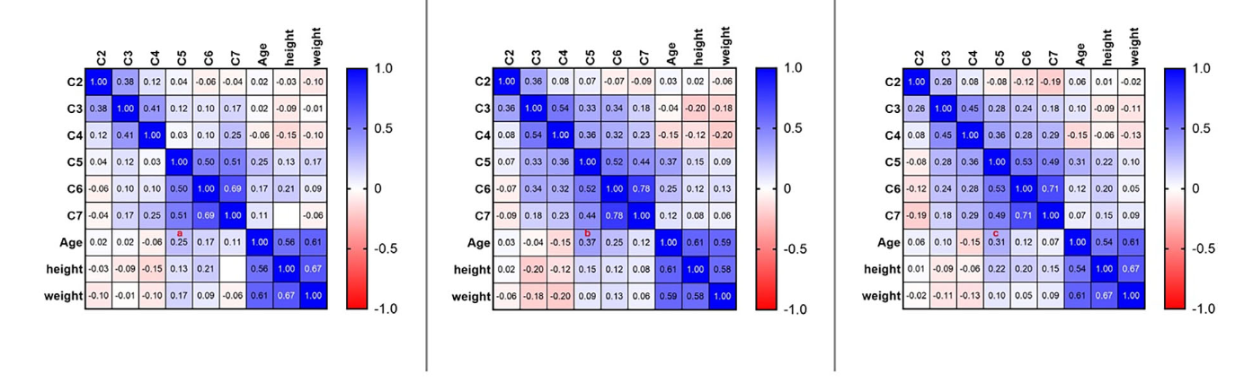
Correlation matrix between MTR and age, height, and weight across vertebral levels. C5 shows a significant positive correlation with age.
Discussion
This study provides the first normative MTR dataset for the pediatric spinal cord and reveals:
- A positive age-MTR relationship, reflecting progressive myelination during growth.
- Sex-based MTR differences, consistent with greater white matter density in females.
- Rostro-caudal MTR decline, likely due to axonal density and white matter volume changes along the spinal cord.
These findings parallel adult diffusion MRI trends, but with distinct developmental patterns in the pediatric cord.
Such normative metrics are essential for interpreting disease-related changes in pediatric SCI, demyelination, or developmental disorders.
Key Takeaways
- MTR increases with age, peaking near the C5 level.
- Females exhibit higher MTR values, suggesting earlier or more pronounced myelination.
- Provides a normative database to compare against pediatric spinal cord pathologies.
- Demonstrates feasibility of atlas-based MT analysis using automated SCT pipelines.
Future Directions
Next steps include:
- Expanding the normative database to thoracic and lumbar levels.
- Integrating multi-parametric MRI (e.g., DTI, NODDI, DKI) for microstructural insights.
- Applying these benchmarks to pediatric SCI and demyelination cohorts to detect early injury biomarkers.
Citation
Naghizadeh Kashani S., Vel I., Sadeghi Adl Z., Shahrampour S., Middleton D., Alizadeh M., Krisa L., Faro S., Tounekti S., Cohen-Adad J., Mohamed F.
“Magnetization Transfer Ratio in the Typically Developing Pediatric Spinal Cord: Normative Data and Age Correlation.”
Journal of Neuroimaging, 2025; 35:e70019.
👉 Read the full paper